Nipple shadows are apparent on approximate 10% of AP/PA views of the chest. Nipple shadows can be seen in patients with different genders and age. When present, they need to be differentiated from more significant intra thoracic pleural or pulmonary nodules.
General characteristics of nipple shadows include some or all of the following:
- Unilateral or bilateral and symmetric
- Caliber of 5 to 15 mm
- Usually round or oval
- Located between anterior 5th and 6th ribs in males and 9th and 10th posterior ribs. May be asymmetric in caliber and location.
- Along the inferior aspect of the female breasts although variable depending on the size of the breasts
- Margin characteristics are variable and often are partial with incomplete and fuzzy margins or radiolucent halo, sharp lateral border and poor medial border
- May not be present on the recent films
- Prominent nipples may be visible on the lateral view of the chest.
If there is doubt whether a nodular opacity represents a nipple shadow or not, a repeat chest x-ray with nipple markers should be performed. Nipple markers can be a useful technique in the evaluation of the nodular densities overlying the expected position of the nipple on a chest radiograph. It is a simple and cost effective tool but at a cost and inconvenience for the patient.
[cta_content id=”19237″]
The presence of the nipple marker density on the nodular shadows usually confirms presence of a nipple shadow. However, there is always the rare occurrence of a real solitary pulmonary or a pleural nodule developing in the region of the expected nipple shadow or being overshadowed by a nipple shadow. A caliber larger than 1.5 cm, irregular margins and shape or asymmetry with opposite nipple shadow if present should be the red flags warranting a further assessment with a CT study. Oblique views of the chest may or may not help.
The nipple marker can be a small metallic marker used in Mammography, BB gun pallet, ball-bearing, unraveled paperclip reformed in a triangle around the nipple or other radio-opaque marker which is stuck to the chest on the nipple. A small punctate metallic marker placed directly on the center of the nipple is preferred. Very large caliber markers could obscure the nodule itself and could cover a very large area beyond the margins of the nipple. When feasible, the markers should be placed by medical personnel to avoid faulty placement of the nipple markers.
Usually a comparable PA view of the chest should suffice. Lateral view could be useful in confirming improper placement of the nipple markers. If the marker does not correspond to the nodule then a CT study is warranted for a possible intrathoracic nodular lesion.
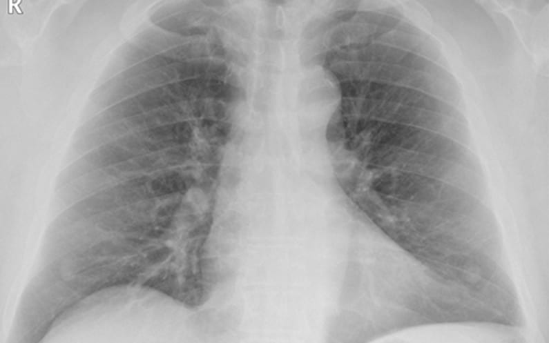
PA VIEW OF THE CHEST WITH NIPPLE MARKERS CONFIRMS BILATERAL NIPPLE SHADOWS
In certain clinical settings it might be useful to place nipple markers on all the patients undergoing a chest x ray examination. Small punctate metallic nipple markers will not obscure other anatomic areas in the chest. This could be a time and money saving tool in emergency rooms and urgent care centers where rapid turnaround time is necessary. Returning patient to the facility for a repeat chest x ray requires facility to contact the patient and then expect the patient to comply. Also, in many clinical settings it may not be possible to contact and return patients to the facility due to improper patient contact information, patients in transit and patient compliance issues.
Get Accurate Overreads Quickly
The teleradiology overread service you choose is an extension of your clinic. Experity offers industry-leading turnaround time, 99.94% accuracy rates, and real-time access to radiologists. With experts by your side – assisted by AI – you ensure better diagnostics and improve outcomes for your patients.